


Advanced Wound Care
We empower healthcare professionals to treat people with wounds, by providing innovative dressings and skin-substitute products, as well as training and support for healthcare professionals.

Siegrid
|
User, Advanced Wound Care
Introduction
Chronic wounds can take years to heal, and for many people living with wounds, it can seriously impact their quality of life. Wounds can present a complex challenge for healthcare professionals: with limited training and without the right products available, wounds are more likely to get infected – prolonging or complicating treatment and healing.
Coloplast has developed a portfolio of advanced wound care products, designed to reduce the risk of infection and help patients heal faster. In 2023, Coloplast acquired Kerecis, a pioneering company that uses intact fish skin technology to help manage hard-to-heal wounds.
Simplifying wound healing
Managing exudate in wounds is key to achieving optimal wound healing conditions. Too much or too little can delay healing and lead to complications like infection. Based on years of clinical research, we’ve developed a range of foam and fiber dressings that help to reduce this risk or treat existing infection.
Our Biatain® foam dressings feature 3DFit Technology, that makes the foam expand when it comes into contact with exudate, matching the shape and depth of the wound*. By creating a close fit to the wound bed, the dressing reduces exudate pooling, lowers the risk of infection and maceration, and leads to faster healing.
Our products

Biatain® Silicone
Made with soft and conformable foam, Biatain® Silicone with 3DFit Technology has a gentle silicone adhesive, a lock-away layer and a semi-permeable, waterproof topfilm. It may be left in place for up to 7 days and can be used on a broad range of acute and chronic wounds – such as venous leg ulcers, pressure ulcers and non-infected diabetic foot ulcers.

Biatain® Fiber
A soft, gelling fiber dressing featuring HexaLock Technology, designed for highly exuding wounds, sloughy wounds, and cavity wounds including undermining.

Comfeel® Plus
Comfeel Plus® is a hydrocolloid dressing that can be used to treat non-to-low exuding wounds in the final stages of healing. It provides ensured protection, wound monitoring, and user-friendly design.
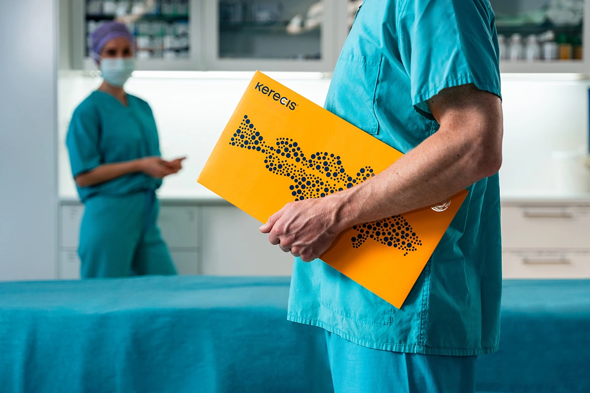
Biologics
Kerecis is pioneering the use of intact fish skin to promote tissue regeneration in chronic and hard-to-heal wounds and burns. We also produce intact fish skin grafts for surgical use.
Structurally closer to human skin than other graft substitutes, fish skin sourced from sustainably harvested North Atlantic waters poses no known risk of viral disease transmission. This means that, unlike human or porcine skin, the fish skin doesn’t have to be treated with harsh detergents. Instead, we gently process the fish skin in a way that preserves the skin’s three-dimensional structure, maintaining its inherent natural strength, complexity, and molecules (such as fatty acids).
Cleared by the FDA and European regulatory authorities for wound management, Kerecis intact fish skin products can be used to treat pressure ulcers, vascular ulcers, diabetic wounds and draining wounds, as well as trauma and surgical wounds.

MariGen® Expanse
- 1. Voegeli D, Landauro MH, Sperup T, Ayoub N, McRobert JW. Clinical performance and cost-effectiveness of a Silicone foam with 3DFit™ technology in chronic wounds compared with standard of care: An open randomised multicentre investigation. Int Wound J. 2024; 21(12).
- *Conformability may vary across product design.
- **Based on UK pricing data.
- ***In wounds up to 2cm in depth. Standard of care: Aquacel® Extra filler combined with a Mepilex® secondary dressing.